De Arachniden - Systematiek
The Arachnids - Systematics
This page is dedicated to Frans
Janssens - Deze pagina is opgedragen aan Frans Janssens
Something
about legs and leg-bearing segments
Iets over poten en
poot-dragende segmenten
GENERAL
REMARKS
Some of the more important
remarks about legs and leg-bearing segments amongst the arthropods may be
enumerated as follows:
1. The generalized type of an
arachnid leg appears to possess one more segment than the maximum number
of eight, but this point needs further investigation. An arachnid leg so
constituted should have the following segments, named from the base to
tip: Subcoxa, coxa, coxal trochanter, femoral trochanter, femur, patella,
tibia, tarsus, and pretarsus.
2. The generalized pauropod leg
is composed of eight rings, which represent, however, only six or possibly
seven true segments, the first three of which probably represent the coxa,
first trochanter, and second trochanter. The last four true segments of
the pauropod leg are the femur, tibia, tarsus, and pretarsus.
3. The generalized symphylid leg
is composed of seven true segments, the first being represented by a
condyle-bearing plate. They are: Subcoxa, coxa, a greatly enlarged
trochanter, a much reduced femur, a tibia, tarsus, and pretarsus.
4. The generalized thysanuran
leg is completely homologous with the pauropod type, with the exception
that it possesses a subcoxa, usually in the form of a platelike structure.
5. The typical collembolan leg
possesses a subcoxal segment and either lacks the tarsus entirely or has
it represented by a short rudiment at the base of the claws.
6. The so-called coxal
appendages in the Pauropoda, Symphyla, and the Thysanura are not true
appendages and have no muscle fibers attaching to them. They are probably
not homologous among themselves. Some may represent structures analogous,
or possibly even homologous, with either the epipods or the exopods of
Crustacea.
7. The primitive insectan type
of tarsus was three-clawed. Two-clawed or one-clawed types found in the
Thysanura or the Collembola are clearly not primitive but evidently were
derived from the three-clawed type as found today in the Pauropoda,
Symphyla, and certain Thysanura.
8. Much evidence exists
indicating the presence of an additional segment to the insect thorax,
which should probably be called the cervical. It should be considered as
being homologous with the legless postcephalic segment of pauropods and
certain symphylids.
9. The so-called intersegmental
region of certain thysanurans should not be regarded either as a distinct
segment or as an intersegmental structure but each region as an integral
part of the segment bearing the adjoining posterior sternite.
10. The primitive thoracic
tergal plates are simple structures without condyles or apodemes and did
not completely cover the dorsal surface of the segments on which they were
situated.
11. The primitive thoracic
sterna of an insect were probably transversely divided into two sternal
plates, the posterior of which articulated laterally with the inner
condyles of the coxae.
12. In general, all vestiges of
pleural plates are wanting in those arthropods which have a cylindrical
and functional subcoxa. As the subcoxa becomes reduced and its inner
(ventral) walls obliterated, two outer crescentic plates persist as body
sclerites. They appear to have resulted from the development of a
pseudosegmentation of the subcoxa. The distal one of these two sclerites
becomes the trochantin of generalized insects and the proximal one the
eupleuron, or " mother " pleural plate from which certain others
were later derived by " fragmentation."
13. The subcoxal theory of the
origin of the pleural plates of insects is sound, and receives further
support .
ALGEMENE
OPMERKINGEN
Enkele van de belangrijkste
opmerkingen omtrent poten en poot-dragende segmenten bij de arthropoden,
kunnen we als volgt opsommen:
1. Het algemene type van een
poot van een arachnide lijkt één segment meer te hebben dan
het maximale aantal van acht, maar dit vraagt verder onderzoek. Een poot
van een arachnide zou de volgende segmenten, benoemd van basis naar top,
moeten hebben:Subcoxa, coxa, coxale trochanter, femorale trochanter,
femur, patella, tibia, metatarsus en tarsus.
2. De typische poot van een
pauropode is samengesteld uit acht ringen, die slechts zes of mogelijk
zeven echte segmenten, waarvan de eerste drie de coxa, eerste trochanter
en tweede trochanter zijn, voorstellen. De laatste vier echte segmenten
van de poot van een pauropode zijn femur, tibia, tarsus en pretarsus.
3. De typische poot van een
symphylide is samengesteld uit zeven echte segmenten, waarvan de eerste
uit een condyle draagplaat bestaat. Ze zijn: Subcoxa, coxa, een enorm
vergrootte trochanter, een veel kleinere femur, een tibia, tarsus en
pretarsus.
4. De typische poot van een
thysanura is volledig homoloog met deze van het pauropode type, met deze
uitzondering dat hij over een subcoxa, gewoonlijk in de vorm van een
plaatachtige structuur, beschikt.
5. De typische collembola-poot
heeft een subcoxaal segment en heeft ofwel geen tarsus of heeft deze
vertegenwoordigd door een kort rudiment aan de basis van de klauwen.
6. De zogenoemde coxale
aanhangels bij de Pauropoda, Symphyla en de Thysanura zijn geen echte
aanhangsels en hebben geen spiervezels eraan vastgehecht. Zij zijn
waarschijnlijk niet onderling homoloog. Sommige kunnen structuren analoog,
misschien zelfs homoloog, vertegenwoordigen, met ofwel de epipoda of the
exapoda van de Crustacea.
7. Het primitieve tarsustype van
de insecten was drieklauwig. Tweeklauwige of éénklauwige
types die bij de Thysanura of de Collembola aangetroffen worden, zijn
duidelijk niet primitief maar zijn waarschijnlijk afgeleid van het
drieklauwige type, zoals dit vandaag wordt aangetroffen bij de Pauropoda,
Symphyla en bepaalde Thysanura.
8. Er bestaan veel bewijzen die
op de aanwezigheid van een bijkomend segment, waarschijnlijk het
cervicale, van de insectenthorax wijzen. Dit zou als homoloog met het
pootloze poscephale segment van pauropoden en bepaalde symphyliden dienen
beschouwd te worden
9. De zogenoemde intersegmentale
streek van bepaalde thysanura mag geenszins als een duidelijk segment of
als een intersegmentale structuur beschouwd worden, maar elke streek als
een essentieel deel van het segment dat de aangrenzende achterste sterniet
draagt.
10. De primitieve thoracale
tergaalplaten zijn eenvoudige structuren zonder condylen of apodemen en
het dorsaal oppervlak van de segmenten waarop zij zich bevinden, niet
volledig.
11. De primitieve thoracale
sterna van een insect waren waarschijnlijk transversaal verdeeld in twee
sternaalplaten, waarvan de achterste lateraal articuleerde met de
binnencondylen van de coxae.
12.Alle restanten van
pleuraalplaten ontbreken in 't algemeen bij deze arthropoden die een
cylindrische en functionele subcoxa hebben. Wanneer de subcoxa gereduceerd
raakt en haar binnen (ventrale) wanden vernietigd zijn, blijven twee
buitenste halfmaanvormige platen over als lichaamssklerieten. Zij lijken
te voort te komen van de ontwikkeling van een pseudosegmentatie van de
subcoxa. De distale van deze twee sklerieten wordt de trochantin van
insecten in 't algemeen en de proximale het eupleuron of de "moeder"-
pleuraalplaat waarvan bepaalde andere later werden afgeleid door "fragmentatie".
13. De subcoxale theorie over de
oorsprong van de pleuraalplaten van insecten is degelijk en krijgt verder
bijval.
USED
ABBREVIATIONS - GEBRUIKTE AFKORTINGEN
| ac |
anterior tarsal claw
voorste tarsale klauw |
map |
medium apodeme
middenste apodema |
| ant |
antenna
antenne |
pat |
patella |
| ap |
coxaal of trochantaal aanhangsel
coxal or trochantal appendage |
pc |
posterior tarsal claw
achterste tarsale klauw |
| bs |
basisternum |
plp |
pleuraalplaat
pleural plate |
| cl |
claw
klauw |
prec |
precosta |
| cx |
coxa |
pt |
pretarsus |
| dc |
depressor of coxa
coxale depressor |
rc |
rotator of coxa
coxale rotator |
| dt |
depressor of trochanter
trochantale depressor |
rt |
rotator of trochanter
trochantale rotator |
| evs |
eversible sac
eversibele zak |
s |
sternum |
| extc |
extensor of claws
klauwextensor |
sI |
eerste lichaamssegment
first body segment |
| extf |
extensor of femur
femurextensor |
scx |
subcoxa |
| extt |
extensor of tibia
tibia-extensor |
sl |
sternellum |
| extp |
extensor of patella
patella-extensor |
sp |
spiracle
spirakel |
| fe |
femur |
sps |
specialized setae
gespecialiseerde setae |
| flc |
flexor of claw
klauwflexor |
stn |
sternite
sterniet |
| flf |
flexor of femur
femurflexor |
ta |
tarsus |
| flp |
flexor of patella
patella-flexor |
terg |
tergite
tergiet |
| flt |
flexor of tarsus
tarsusflexor |
thI |
prothorax |
| fltb |
flexor of tibia
tibiaflexor |
ti |
tibia |
| h |
head
kop |
tr |
trochanter |
| intl |
intersegmental lobe of neck
intersegmentale neklob |
trI |
first trochanter
eerste trochanter |
| l |
leg
poot |
trII |
second trochanter
tweede trochanter |
| lI |
leg I
poot I |
un |
unguiculus
|
| lap |
lateral apodeme
lateraal apodema |
utr |
unguitractor plate
unguitractor-plaat |
| lc |
levator of coxa
coxale levator |
x |
Y-shaped sternal ridge
Y-vormige sternaalkam |
| ls |
laterosternum |
y |
sternal rod
sternaalstang |
| lt |
levator of trochanter
trochantale levator |
|
|
AFBEELDINGEN
- IMAGES
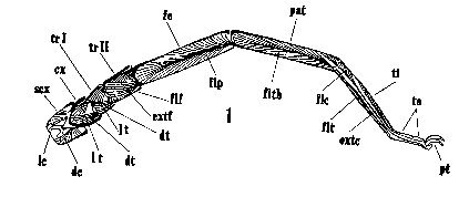
The third left leg of a solpugid, showing muscle attachments
De derde poot van een Solpugida; spieraanhechtingspunten |
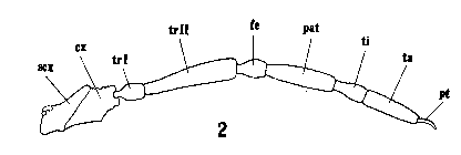
Leg 1 of the primitive phalangid, Holosiro acaroides, with
segments labeled.
Poot 1 van een primitieve hooiwagen (Opiliones), Holosiro acaroides,
met gelabelde segmenten |
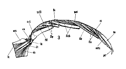
. Front leg of Labidostomma sp. showing muscle attachments
Voorpoot van Labidostomma spec.met spieraanhechtingspunten |
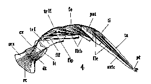
Third leg of the gamasid mite, Macrocheles tridentifer, showing
muscle attachments.
Derde poot van de gamaside mijt Macrocheles tridentifer met
spieraanhechtingen |
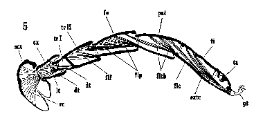
. Front leg of tick, Rhipicephalus sanguineus, showing muscle
attach- ments.
Voorpoot van teek, Rhipicephalus sanguineus, met
spieraanhechtingen |
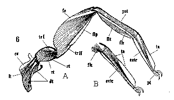
A, hind leg of pseudoscorpion, Chelifer cancroides, showing
muscle attachments; B, end of hind leg of pseudoscorpion, Obisiuim
maritimum, showing same.
A. Achterpoot van pseudoschorpioen, Chelifer cancroidesmet
spieraanhechtingen, B, einde van achterpoot van pseudoschorpioen, Obisiuim
maritimum met spieraanhechtingen |
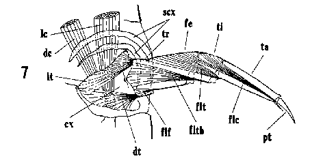
The second leg of Acerentulus barberi showing segmentation and
musculature.
De tweede poot van Acerentulus barberi met segmentatie en
spieren |
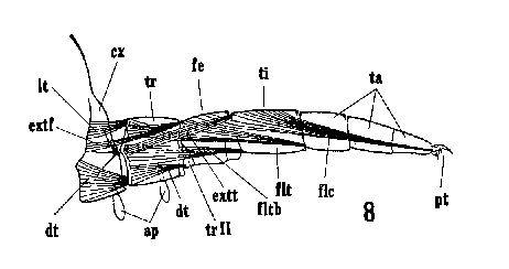
Leg IV of Pauropus sp. showing segmentation and musculature
Poot IV van Pauropus sp. met segmentatie en spieren |
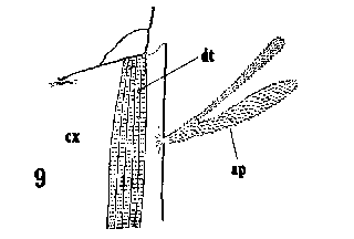
Coxal appendage of last pair of legs of Pauropus sp.
Coxaal aanhangsel van het laatste potenpaar van Pauropus sp. |
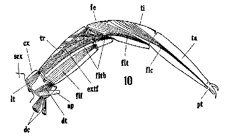
Antepenultimate leg of symphylid, Sculigerella imntaculata,
showing segmentation and muscles
Op twee na de laatste poot van de symphylide Sculigerella
imntaculata met segmentatie en spieren |
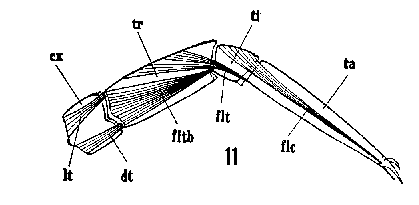
Front leg of a symphylid of the genus Hanseniella showing
segmentation and musculature.
Voorpoot van een symphylide van het genus Hanseniella met
segmentatie en spieren |
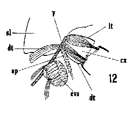
The region about the coxa in a symphylid of the genus Hanseniella,
showing muscle attachments
De coxa-streek van een symhylide van het genus Hanseniella met
spieraanhechtingen |
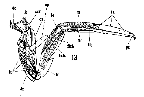
Third leg of Machilis sp. showing segmentation and muscles
Derde poot van Machilis sp. met segmentatie en spieren |

Middle leg of lepismid, Thermobia sp., showing segmentation
Middenpoot van een lepismide, Thermobia sp. met segmentatie |
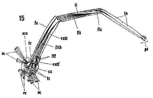
Second leg of Campodea sp., showing segmentation and muscles.
Tweede poot van Campodea sp. met segmentatie en spieren |

Third leg of the collembolan, Tomocerus arcticus, showing
musculature
Derde poot van de collembola Tomocerus arcticus met spieren |
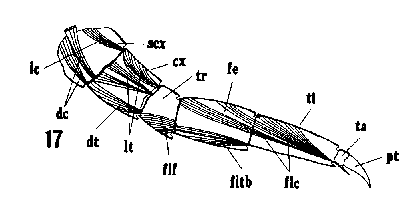
The musculature of third leg of the collembolan, Aphorura ambulans
(Linn.).
Spieren van de derde poot van de collembola Aphorura ambulans (Linn.) |
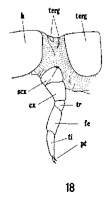
Prothorax, with attached leg, and neck region of collembolan (Isotoma
sp.). Specimen cleared with KOH and stained with acid fuchsin.
Prothorax, met poot, en nekstreek van collembola (Isotoma sp.).
Specimen geklaard met KOH en gekleurd met zure fuchsine. |
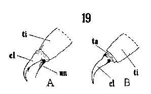
The two types of tarsi found in the generalized Collembola of the
family Poduridae: A, Achorutes; B, Podura.
Twee tarstypen aangetroffen bij de Collembola van de familie Poduridae:
A, Achorutes; B, Podura |
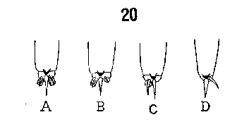
The evolution of the two-clawed tarsus in Pauropus and its
comparison with the two-clawed tarsus in Symphylella. A, B &
C; tarsi I, III and last tarsus of Pauropus; D, tarsus of Symphylella.
(All views from above.)
De evolutie van de tweeklauwige tars bij Pauropus in vergelijk
met de tweeklauwige tars van Symphylella . A,B en C; tarsi I,
III en laatste tars van Pauropus; tars van Symphylella.
(Bovenaanzichten) |
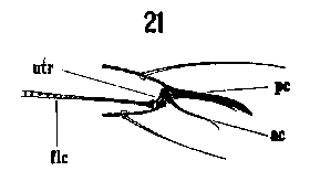
Vertical "optical section" of tip of tarsus I of proturan,
Acerentulus barberi.
Verticale optische doorsnede van het topje van tars I van de protura
Acerentulus barberi |
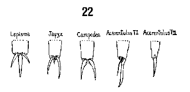
The evolution of the two-clawed type of tarsus in Thysanura and the
one-clawed type in Protura.
De evolutie van het tweeklauwige tars-type bij Thysanura en het éénklauwige
bij Protura |
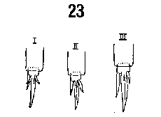
Dorsal view of feet of collembolan, Tomocerus arcticus, showing
variation in claws; I, II, III=tarsi I, II and III respectively.
Dorsaal aanzicht van voetje van de collembola Tomocerus arcticus
met de klauwvariatie; I,II,II = respectievelijk tarsi I, II en III |
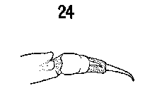
Two-clawed tarsus of isopod crustacean of family Oniscidae.
Tweeklauwige tars van de isopode kreeftachtige van de familie Oniscidae |
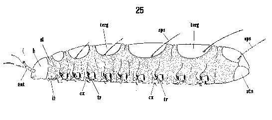
Lateral view of Pauropus huxleyi showing body segmentation,
chitinized parts and specialized setae.
Zijaanzicht van Pauropus huxleyi met lichaamsegmentatie,
gechitiniseerde delen en gespecialiseerde setae |
|
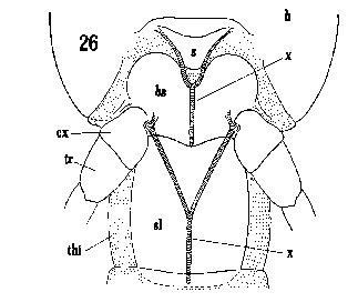
Prothorax and neck region of Japyx bidentatus.
Prothorax en nekstreek van Japyx bidentatus |
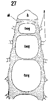
Dorsal view of first six segments of body of Pauropus sp.
showing chitinous plates.
Dorsaal aanzicht van de eerste zes segmenten van het lichaam van Pauropus
sp. met chitineuze platen |
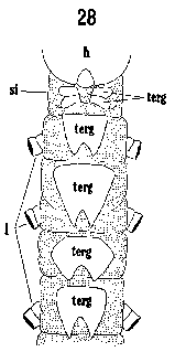
Dorsal view of first five segments of body of Symphylella sp.,
showing chitinous parts.
Dorsaal aanzicht van de eerste vijf segmenten van het lichaam van Symphylella
sp. met chitineuze delen |
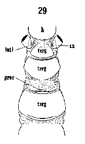
Dorsal view of thorax of Acerentulus barberi showing chitinous
parts.
Dorsaal aanzicht van de thorax van Acerentulus barberi met
chitineuze delen. |
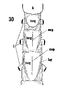
Dorsal view of thorax of Japyx isabellae showing sclerites
Dorsaal aanzicht van Japyx isabellae met sklerieten |
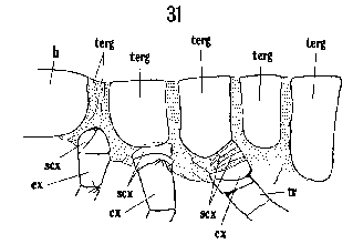
Lateral view of thorax of a springtail, Isotoma sp., together
with proximal parts of legs.
Zijaanzicht van de thorax van een springstaart, Isotoma sp.,
samen met proximale delen van de poten |

Ventral view of cephalothorax of Holosiro acaroides.
Ventraal aanzicht van de cephalothorax van Holosiro acaroides |
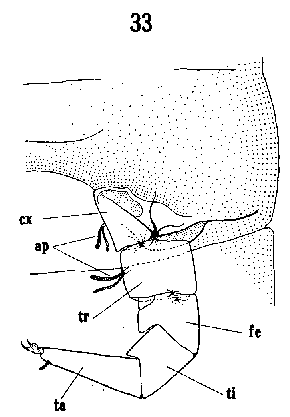
Ventral view of last leg of left side of Pauropus sp. and the
sclerites of body segment it is attached to.
Ventraal aanzicht van de laatste poot van de linkerzijde van Pauropus
sp. en de sklerieten van het lichaamssegment waaraan deze bevestigd is. |
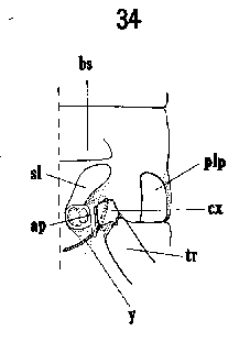
Ventral view of left half of body segment of Scutigerella sp.
showing chitinizations
Ventraal aanzicht van de linkerhelft van het lichaamssegment van Scutigerella
sp. met chitinisaties |
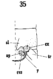
Ventral view of left half of body segment of Hanseniella sp.
showing chitinizations.
Ventraal aanzicht van de linkerhelft van het lichaamssegment van
Hanseniella sp. met chitinisaties |
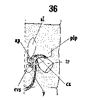
Ventral view of left half of body segments of Symphylella sp.
showing chitinizations.
Ventraal aanzicht van de linkerhelft van het lichaamssegment van Symphylella
sp. met chitinisaties |
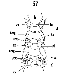
Ventral view of thorax of proturan, Acerentulus barberi.
Ventraal aanzicht van protura Acerentulus barberi |
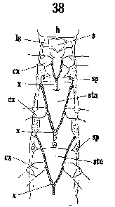
Ventral view of thorax of Japyx isabellae, showing sclerites.
Ventraal aanzicht van Japyx isabellae met sklerieten |
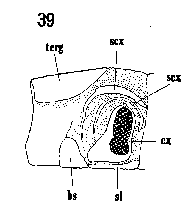
Lateral view of left side of mesothorax of Acerentulus barberi.
Zijaanzicht van de linkerzijde van de mesothorax van Acerentulus
barberi |
|
|
|
|
After H.E.
Ewing "The legs and leg-bearing segments of some primitive arthropod
groups, with notes on leg-segmentation in the Arachnida", Smithsonian
Institution 1928

Deze pagina's © 1999 Gie Wyckmans & Dragon Research&Development
This pages © 1999 Gie Wyckmans & Dragon Research&Development


